Early Signs of Inflammatory Breast Cancer
Regional Cancer Care Associates (RCCA) provides comprehensive treatment of cancer and blood disorders to patients throughout New Jersey, Connecticut, and the Washington, DC, area. Here,
HIPAA Alert: Potential Data Breach Learn More
Questions on Oncology, Hematology and/or Infusion Clinical Services due to COVID-19 Crisis – CALL 833-698-1623
Important Information for Our Patients Regarding the Coronavirus.
RCCA Providing Area Cancer Patients with Access to Care During Coronavirus Outbreak
RCCA Offering Patients Virtual Visits During Coronavirus Pandemic
When it comes to treating breast cancer, early detection is the first line of defense. The sooner cancer is diagnosed, the more treatable it is. Routine mammograms play an important part in this. Most new cancer cases are diagnosed through annual mammogram testing. This allows treatment to begin long before cancer creates a noticeable lump, giving patients an excellent chance of recovery.
Many individuals who are diagnosed with breast cancer or other cancer types turn to Regional Cancer Care Associates (RCCA), which features more than 100 medical oncologists and hematologists who serve patients in 25 locations throughout New Jersey, Connecticut, Massachusetts, and the Washington, DC area. Following is a discussion on breast cancer and the importance of early detection.
When you are diagnosed with breast cancer and require surgical treatment, you may undergo a mastectomy, the removal of some or all of the breast. There are several types of mastectomy procedures, some of which may involve only the removal of lymph nodes, muscle tissue and skin depending on which mastectomy you and your oncologist decide on.
Here is a breakdown of the types of mastectomy surgeries performed at Regional Cancer Care Associates.
Breast cancer is one of the most common cancers in the United States, and the second most common cause of cancer death in women. According to studies by American Cancer Society, about 316,950 new cases of invasive breast cancer are diagnosed each year. One in eight women, or 13%, will develop breast cancer at some point.
Fortunately, breast cancer death rates have been consistently declining since 1989. Part of this decline is due to cancer research uncovering new and improved treatments. More significant, however, has been the implementation of routine screening. Frequent mammograms make early detection possible, allowing for prompt treatment with better outcomes.
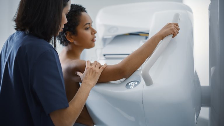
Mammograms are X-ray images of the breasts. They are used to non-invasively examine the breast’s tissue and internal structures to check for early signs of cancer. Mammograms may be taken as part of routine screening, or they may be used to diagnose the cause of an individual’s symptoms.
While medical research has led to improved breast cancer treatments, early detection provides the best outlook. In breast cancer stages 0 and I, survival rates are between 98% and 100%. This rate decreases as cancer progresses into its later stages. In Stage IV, treatment usually focuses on symptom management. This is why it’s important to diagnose cancer early, while it is most treatable.
Routine mammograms make early diagnosis possible. Breast cancer appears on mammograms, often before it causes symptoms or a noticeable lump. Treatment can begin earlier, giving the patient a better chance of a positive outcome.
Routine mammograms are simple procedures. The entire process takes only a few moments and is painless, though it may cause some discomfort. Individuals can expect:
This process is repeated four times, to get a top view and a side view of each breast. Afterward, the individual may need to wait while the technologist checks the X-rays to ensure the pictures are high-quality for analysis. If the pictures are incomplete, out of focus, or otherwise flawed, they may need to be redone. If the pictures are satisfactory, they are sent to the radiologist for analysis.
If an individual is recommended for testing because breast cancer is already suspected, they receive what is called a diagnostic mammogram. This uses the same equipment and follows the same process as a routine mammogram but may take slightly longer as more pictures are taken. The additional pictures allow the radiologist to take a closer look at suspicious areas to confirm the presence of cancer.

Once a mammogram produces satisfactory images, they are sent to a radiologist. This is a medical professional with special training in analyzing X-ray images. The radiologist examines the mammograms closely for signs of cancer, which may include:
Abnormalities detected in mammograms are not always evidence of cancer. Calcifications can be caused by other conditions, and a mass may be a cyst or non-cancerous tumor like fibroadenoma. If a mammogram reveals an abnormality, further testing is needed to determine if it is cancer.
If a mammogram reveals an abnormality, the medical oncologist will request further tests. These will help the oncologist determine whether the abnormality is caused by cancer and learn more about its characteristics. Every case of breast cancer is unique. The more oncologists understand about a person’s case, the better they can plan an effective treatment.
Ultrasound is another imaging technology used to diagnose conditions in the breast. Instead of taking an image of the full breast, as with a mammogram, ultrasound technologists more frequently focus on the suspicious area. This allows for a closer look at the abnormality for more detailed analysis.
Magnetic resonance imaging (MRI) is also sometimes used to diagnose breast cancer. This technology uses radio waves and powerful magnets to create images. It may be used to determine a tumor’s exact location or to look for other tumors in the breast.
A biopsy involves removing a small amount of tissue or fluid from the abnormality. This sample is tested in a laboratory to determine whether the abnormality is cancerous. It can also determine properties such as responsiveness to hormones or the presence of certain proteins that may be used for treatment.

Mammograms are the most reliable way to detect breast cancer early for a successful treatment. A person’s first mammogram can be an anxiety-filled event. However, you can alleviate these feelings by becoming educated and knowledgeable about the screening procedure and what it involves.
Breast cancer and other cancer types are treated at Regional Cancer Care Associates (RCCA), a group of more than 100 medical oncologists and hematologists who provide care to more than 30,000 new patients and 265,000 established patients each year. RCCA offers some of the latest therapies, including targeted treatments like immunotherapy or hormone therapy. Patients also have access to cutting-edge diagnostics and clinical trials in community-based centers close to home.
RCCA recommends that individuals start receiving annual mammograms starting at age 40. People who are at risk of breast cancer may need to start sooner or be screened more frequently. Talk with your healthcare provider about recommended mammogram frequency for your level of risk.
Getting a mammogram takes only a few moments and is painless.
Radiologists look for anything that appears abnormal or unlike other breast tissue. Abnormalities include white spotting caused by calcifications, defined tissue masses, changes in the tissue pattern, or distortion of the overall breast structure.
No, abnormalities on a mammogram are not always cancer. They may be caused by other conditions, past injuries, or the positioning of the breast between the plates. If the radiologist finds anything suspicious, further testing is needed to determine whether it is cancer.
The prospect of mastectomy can be both frightening and heartbreaking, but you won’t be in this fight alone. Our team of experts at Regional Cancer Care Associates will work tirelessly to form an appropriate course of action to battle your breast cancer. We will review the types of mastectomy that may be an option for you and prepare you for what to expect before, during and after the procedure. We will also support you long after your mastectomy to ensure your healing process is successful, from breast reconstruction surgery to follow-up visits once you’re completely cancer-free. For more information, contact Regional Cancer Care Associates today.

Regional Cancer Care Associates (RCCA) provides comprehensive treatment of cancer and blood disorders to patients throughout New Jersey, Connecticut, and the Washington, DC, area. Here,

When people think of breast cancer, they generally think of it affecting women. However, in rare circumstances, breast cancer can affect men, most commonly in

First diagnosed with breast cancer in 1992, she has persevered in her battle against the disease for more than 30 years.

Regional Cancer Care Associates is one of fewer than 200 medical practices in the country selected to participate in the Oncology Care Model (OCM); a recent Medicare initiative aimed at improving care coordination and access to and quality of care for Medicare beneficiaries undergoing chemotherapy treatment.