Why Get Called Back After a Mammogram?
It is not unusual for individuals to be recalled after an initial mammogram screening. The recall is not always due to suspicious results. There are many possible reasons for a radiologist to call the individual back, including:
- The first images were unclear
- The first images missed part of the breast tissue
- There is an area that looks different from the rest of the breast
- The mammogram revealed a suspicious mass or calcification
Being recalled may cause someone to worry. However, fewer than 1 in 10 women are actually found to have cancer after being called back. In most cases, suspicious findings no longer look unusual under close examination. The follow-up provides certainty that the finding is benign. If so, the individual can enjoy peace of mind that they are truly cancer-free. If screening does reveal a problem, then treatment can begin as soon as possible for the best chances of success.
Understanding Abnormal Mammogram Results
When discussing next steps after an abnormal mammogram, it helps to understand what the results mean. Physicians use the Breast Imaging Reporting and Data System (BI-RADS) to describe mammogram findings. Categories run from zero to six:
Category 0
The findings are incomplete. Additional imaging is needed to clarify the results. The radiologist may request another mammogram with a different view, one with spot compression, or an ultrasound.
Category 1
The findings are negative. The mammogram reveals nothing new or abnormal. The individual’s breasts are symmetrical with no masses, unusual tissue structures, or calcifications.
Category 2
The findings are benign and non-cancerous. This may include masses, lymph nodes, or calcifications. Calcifications are calcium deposits that can be seen on mammograms but are typically too small to be felt in breast tissue. They are a byproduct of breast cell growth and division. There are two types:
- Microcalcifications: These are tiny calcifications that may require further testing, as they can become cancerous.
- Macrocalcifications: These are large, coarse deposits that are commonly found in women age 50 and older. They are the natural result of aging, past injuries, or buildup.
Category 2 results are negative for cancer. Noting them, however, is important to inform radiologists for future mammogram screenings.
Category 3
The findings are probably benign. Findings in this category have less than a two percent chance of being cancerous and are not expected to become cancerous. However, they cannot be definitively labeled benign. Prompt follow-up is required in order to conclusively rule out cancerous results. In most cases, the radiologist will suggest further imaging within six to 12 months, repeated regularly over two years, or until the finding is determined to be stable.
Category 4
The findings are suspicious or abnormal. They are possibly, but not definitely, cancerous. There are three subcategories based on the estimated likelihood of cancer:
- 4A: The likelihood of cancer is low, between 2% and 10%.
- 4B: The likelihood of cancer is moderate, between 10% and 50%.
- 4C: The likelihood of cancer is high, between 50% and 95%.
A surgical biopsy is recommended after a Category 4 finding to confirm the presence or absence of cancer. Biopsy involves removing a small amount of breast tissue for evaluation.
Category 5
The findings are highly likely to be cancer. Findings look like cancer and are at least 95% likely to be cancer. A surgical biopsy is very strongly recommended.
Category 6
The findings are definitely cancerous. This category is only used after the presence of cancer is proven by a biopsy. At this point, further imaging is used to monitor how the cancer responds to treatment.
What Happens at a Mammogram Follow-Up Appointment?
If routine mammogram findings are Category 2 or higher, a follow-up appointment will be made. This typically involves another mammogram called a diagnostic mammogram. Rather than taking images of the whole breast, the radiologist will take several views of a particular part of the breast.
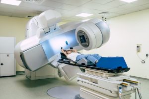
The radiologist may also request other types of medical imaging. Ultrasound uses sound waves to get images and may provide a better view of structures that were unclear on the mammogram. Magnetic resonance imaging (MRI) uses radio waves and magnets. MRI images are far more detailed than mammograms or ultrasounds and can detect very tiny changes in breast tissue.
If the area is still suspicious after detailed imaging, a biopsy will be performed. This procedure uses a hollow needle to take small samples of tissue from the breast. The samples are then examined under a microscope. Biopsy is the only way to be sure whether a suspicious growth is cancerous.
Mammograms and Dense Breast Tissue
Additional testing is often required for women with dense breasts. This is because dense breasts are more difficult to evaluate with mammograms. They have a high composition of glandular and fibrous tissue, which can disguise abnormal growths and make mammograms more difficult to interpret. Breast density is recorded in four categories:
- Category A: The breasts are largely fatty.
- Category B: The breasts feature scattered areas of dense tissue.
- Category C: The breasts feature an even (heterogenous) distribution of dense tissue.
- Category D: The breasts are extremely dense.
Dense breasts are a normal, common occurrence. Almost half of women age 40 and older have them. These women may be asked for follow-up imaging more often than others, but not because they are more likely to have cancer. This is only to ensure imaging results are clear.
Find Breast Cancer Care Near You in NJ, CT, MA, and the Washington, D.C., Area
Waiting for an abnormal mammogram follow-up can be unnerving. If someone feels nervous, they can speak with loved ones who have had screenings or biopsies. They can also contact a physician to learn more about breast cancer and test recommendations. Regional Cancer Care Associates educates patients to help them avoid life-threatening cancer. RCCA specialists provide care to more than 30,000 new patients and 265,000 established patients each year. RCCA physicians offer patients innovative therapies, including immunotherapies and targeted therapy, cutting-edge diagnostics as well as access to approximately 300 clinical trials in community-based centers close to home.
Frequently Asked Questions About Breast Cancer Screening
Why are routine mammograms important?
Routine mammograms can detect signs of cancer before an individual notices symptoms or a lump. This allows treatment to begin early, giving patients a good chance for full recovery.
Are mammograms safe?
Mammograms use only small doses of radiation. The risk of harm is very low. Repeated mammograms can increase cancer risk, however, so women should speak with their doctors about what is suitable, especially if there is a possibility of pregnancy.
How often should I get routine mammograms?
Most authorities recommend annual mammograms starting after age 40. High-risk women may begin mammograms earlier or supplement screenings with additional tests, such as MRI scans.
Does dense breast tissue increase cancer risk?
Dense breasts do slightly increase a person’s risk of developing cancer. They also make it harder to read mammograms.